

Bladder cancer is a disease in which malignant cells arise in the bladder. Symptoms can include blood in the urine, pain during urination, increased frequency of passing urine, or feeling the need to urinate but with nothing coming out. The bulk of bladder cancers are histlogically classed as transitional cell carcinomas which arise in the uroepithelium (lining of the bladder). Other types include squamous cell carcinomas, and adenocarcinomas. Treatment will depend on how far the tumour has invaded the surrounding tissues, and if it has spread to other parts of the body. World-wide about 260,000 people are diagnosed with bladder cancer each year.
Saturday, October 17, 2009
Wednesday, September 16, 2009
Lung cancer
Lung cancer is a disease of uncontrolled cell growth in tissues of the lung. This growth may lead to metastasis, which is the invasion of adjacent tissue and infiltration beyond the lungs. The vast majority of primary lung cancers are carcinomas of the lung, derived from epithelial cells. Lung cancer, the most common cause of cancer-related death in men and also the most common in women, is responsible for 1.3 million deaths worldwide annually. The most common symptoms are shortness of breath, coughing (including coughing up blood), and weight loss.
The main types of lung cancer are small cell lung carcinoma and non-small cell lung carcinoma. This distinction is important, because the treatment varies; non-small cell lung carcinoma (NSCLC) is sometimes treated with surgery, while small cell lung carcinoma (SCLC) usually responds better to chemotherapy and radiation. The most common cause of lung cancer is long-term exposure to tobacco smoke. The occurrence of lung cancer in nonsmokers, who account for as many as 15% of cases , is often attributed to a combination of genetic factors, radon gas, asbestos, and air pollution, including secondhand smoke.
Lung cancer may be seen on chest radiograph and computed tomography (CT scan). The diagnosis is confirmed with a biopsy. This is usually performed via bronchoscopy or CT-guided biopsy. Treatment and prognosis depend upon the histological type of cancer, the stage (degree of spread), and the patient's performance status. Possible treatments include surgery, chemotherapy, and radiotherapy. With treatment, the five-year survival rate is 14%.
Source : wikipedia.org
Source : wikipedia.org
Tuesday, September 8, 2009
 |  |  | |
- A to Z List of Cancers
- Are Your Symptoms Recurrent? Identify Your Genital...
- The Sunshine Vitamin: Vitamin D And Your Health
- Build Bone Density With Osteoporosis Prevention!
- The most common sexually transmitted disease in th...
- HPV Test: The Best Method In Determining The Cervi...
- Rates Of Cervical Cancer Rising Among Teens
- Study Says Few Birth Control Pills Are Safe
- Tips For Women To Deal With Endometriosis Pain
- Tiny Ovarian Tumors Hard To Detect
- Cone Biopsy – Best Method In Detecting Cervical Di...
- Women Who Have Had Breast Cancer Should Avoid Alco...
- HIV, National Women And Girls Awareness Day

Monday, September 7, 2009
Symptoms of mesothelioma
Mesothelioma often starts as a lot of tiny lumps (nodules) in the pleura, which may not show up on scans or x-rays until they are quite large. The main symptoms of pleural mesothelioma are breathlessness and chest pain. Some people find that their voice becomes hoarse and they have a cough that does not go away.
Peritoneal mesothelioma often causes swelling and pain in the abdomen.
General symptoms
Both types of mesothelioma can cause other general symptoms, such as loss of appetite, sweating (especially at night), weight loss and tiredness. As many of these symptoms can also be caused by other illnesses, your doctor will need to do a series of tests before a diagnosis can be made.
Causes of mesothelioma
Asbestos is the most common cause of mesothelioma. Up to nine out of ten cases of mesothelioma are caused by exposure to asbestos. Asbestos is a natural mineral, mined from rock found in many countries. It is made up of tiny fibres that are as strong as steel but can be woven like cotton and are highly resistant to heat and chemicals.
During the 1960s the first definite link between mesothelioma and asbestos was made. In the past asbestos was imported to the UK in large quantities. It was used in construction, ship-building and in household appliances. Asbestos was very widely used in insulation materials, such as amosite insulation board, and building materials, including asbestos cement.
When asbestos is disturbed or damaged, it releases tiny fibres that can be breathed into the lungs. Asbestos fibres are very fine and, when breathed in, they can make their way into the smallest airways of the lung, so they cannot be breathed or coughed out. Once the fibres are in the lungs, the body's defence mechanism tries to break them down and remove them, which leads to inflammation in the lung tissue.
The asbestos fibres can also penetrate through the lung tissue to settle in the pleura (the membrane around the lung). Over many years they can cause mesothelioma or other lung diseases to develop.
Asbestos fibres can also be swallowed, and some of the fibres can stick in the digestive system. They can then move into the membrane that lines the abdomen (the peritoneum), where they cause inflammation.
The people most likely to have been exposed to asbestos include:
- construction workers
- plumbers
- electricians
- boilermakers
- shipbuilders
- demolition workers
- people who worked in other places where asbestos was present and
- people who lived near to asbestos factories.
Family members of people who worked with asbestos and brought the dust home on their clothes have also sometimes developed mesothelioma.
There are three types of asbestos: blue, brown and white. Blue and brown asbestos are the types most commonly linked with mesothelioma. They are now very rarely used and cannot be imported into the UK. Originally, white asbestos was thought not to be dangerous but recent studies have now shown that it is also harmful.
In the 1980s, imports of blue and brown asbestos into the UK were stopped, and in 1999 the importation and use of all asbestos was banned. However, as mesothelioma develops so slowly, it is estimated that by 2015 approximately 3000 people will be diagnosed with mesothelioma each year. The number of people who develop mesothelioma will then start to reduce each year.
Mesothelioma does not usually develop until many years after exposure to asbestos. It can take any time from 10 to 60 years, although the average is about 30 to 40 years after exposure to asbestos.
Occasionally, mesothelioma develops in people who have never been exposed to asbestos. The other causes of the disease are not fully understood, but in rare cases the development of mesothelioma has been linked to exposure to radiation.
Research has not found any evidence that smoking increases a person's risk of developing mesothelioma. It is also thought that exposure to other building materials such as fibreglass does not increase the risk.
Mesothelioma is not contagious and cannot be passed on to other people. It is not caused by inherited faulty genes and so family members do not have an increased risk of developing it, unless they have been in contact with asbestos.
What is cancer?
The organs and tissues of the body are made up of tiny building blocks called cells. Cancer is a disease of these cells.
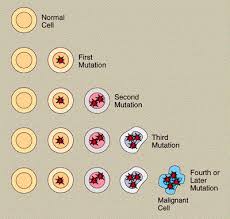
Cells in different parts of the body may look and work differently but most reproduce themselves in the same way. Cells are constantly becoming old and dying, and new cells are produced to replace them. Normally, cells divide in an orderly and controlled manner. If for some reason the process gets out of control, the cells carry on dividing, developing into a lump which is called a tumour.

Tumours can be either benign or malignant. Cancer is the name given to a malignant tumour. Doctors can tell if a tumour is benign or malignant by examining a small sample of cells under a microscope. This is called a biopsy.
In a benign tumour the cells do not spread to other parts of the body and so are not cancerous. However, if they continue to grow at the original site, they may cause a problem by pressing on the surrounding organs.
A malignant tumour consists of cancer cells that have the ability to spread beyond the original area. If the tumour is left untreated, it may spread into and destroy surrounding tissue. Sometimes cells break away from the original (primary) cancer. They may spread to other organs in the body through the bloodstream or lymphatic system.
The lymphatic system is part of the immune system - the body's natural defence against infection and disease. It is a complex system made up of organs, such as bone marrow, the thymus, the spleen, and lymph nodes. The lymph nodes (or glands) throughout the body are connected by a network of tiny lymphatic ducts.
When the cancer cells reach a new area they may go on dividing and form a new tumour. This is known as a secondary cancer or metastasis.
It is important to realise that cancer is not a single disease with a single type of treatment. There are more than 200 different kinds of cancer, each with its own name and treatment.
What is mesothelioma?
Mesothelioma is a cancer of the mesothelium. The mesothelium is a thin membrane that lines the chest and abdomen and surrounds the organs in these areas. The lining around the lungs is called the pleura and in the abdomen it is known as the peritoneum.
About 2000 people in the UK are diagnosed with mesothelioma each year.
Mesothelioma of the lining of the lungs, known as pleural mesothelioma, is much more common than mesothelioma in the peritoneum. For every one person with peritoneal mesothelioma, there will be about 12 people who have pleural mesothelioma.
Pleural mesothelioma
The pleura has two layers: the inner (visceral) layer, which is next to the lung; and the outer (parietal) layer, which lines the chest wall. The two layers of the pleura are usually in contact and slide over each other as we breathe. The membranes produce fluid, which allows them to slide over each other easily.
When mesothelioma develops in the pleura (pleural mesothelioma), the delicate membranes thicken and may press inwards on the lung. Fluid may also collect between the two layers of the pleura: this is known as a pleural effusion.

Structure of the lungs and pleura

Peritoneal mesothelioma
The lining of the abdomen is known as the peritoneum. It also has two layers: the inner (visceral) layer, which is next to the abdominal organs, and the outer (parietal) layer, which lines the abdominal wall.
If the mesothelioma is in the peritoneum it is called peritoneal mesothelioma and causes thickening of the membranes surrounding the abdominal organs and a collection of fluid in the abdomen. The collection of fluid is called ascites and causes swelling of the abdomen.
Side view of the abdomen. The peritoneum is shown as the thick line surrounding the abdominal organs.
The Different Types of Cancer
The Different Types of Cancer Include:
Carcinomas: The most common type of cancer, these tumors arise from the cells that cover external and internal body surfaces. The most frequent cancers of this type in the United States are lung, breast, colon, and prostate cancer.
Sarcomas: Cancers that arise from cells found in the supporting tissues of the body, such as bone, cartilage, fat, connective tissue, and muscle.
Lymphomas: Cancers that arise in the lymph nodes and tissues of the body's immune system.
Leukemias: Cancers of the immature blood cells that grow in the bone marrow and tend to accumulate in large numbers in the bloodstream.
The place where a cancer starts is called the primary site. From there, it can spread (metastasize) to other parts of the body. Regardless of where a cancer may spread, it is always named for the place it began. For instance, breast cancer that spreads to the liver is still called breast cancer, not liver cancer.
Different types of cancer can behave very differently. For example, lung cancer and breast cancer are very different diseases. They grow at different rates and respond to different treatments. That is why people with cancer need treatment that is aimed at their particular kind of cancer.
Not all tumors are malignant (cancerous). Benign, or noncancerous, tumors do not spread to other parts of the body and, with very rare exceptions, are not life-threatening.
During the second half of the 20th century, scientists uncovered many of the intricacies of cancer and developed the technology to pinpoint the exact site of the damage to a specific gene, which has had a tremendous impact on the types of therapies now available.
Monday, August 17, 2009
Chemotherapy
Chemotherapy is the treatment of diseases such as cancer with drug therapy. Since the 1960's the development and use of drugs has significantly improved the prognosis for some types of cancer. Chemo- means chemicals, for most types of cancer chemotherapy will consist of a number of different drugs, this is known as combination chemotherapy. Chemotherapy may be given in a variety of ways; Intravenously (IV) -into a vein is the most common, Intramuscularly (IM) -injection into a muscle, Orally -by mouth, Subcutaneously (SC) -injection under the skin, Intralesionally (IL) directly into a cancerous area, Intrathecally (IT)-into the fluid around the spine, or Topically -medication will be applied onto the skin.
Friday, August 14, 2009
Are Your Symptoms Recurrent? Identify Your Genital Herpes Symptoms!
 Genital herpes is a common sexual transmitted disease.
Genital Herpes is an infection of the genitals caused by the herpes simplex virus (HSV).
Actually, HSV-1 And HSV-2 are two types of the herpes simplex virus that affects the genitals.
Mostly, HSV-1 cause herpes on the face and HSV-2 causes genital herpes. This virus, usually, enters your body via small breaks in your skin or mucous membranes.
Most of you, usually, when infected with genital herpes will unaware of the virus presence in the body, because you often experience no genital herpes symptoms or mild symptoms that too unnoticeable.
No matter whether you exhibit mild genital herpes symptoms or no symptoms, you are likely to pass the virus to your sexual partner.
In general, the herpes symptoms are known as outbreaks. If genital herpes symptoms develop then they differ greatly from person to person. Normally, the genital herpes symptoms will develop between 2-7 days after exposure to the virus.
Some times, you cannot experience the genital herpes symptoms until months or years after being infected to the simplex virus.
Mostly, the genital herpes infections appear as small blisters or ulcers on the genitals. In general, the first occurrence of genital herpes is known as primary infection. During the primary infection, you may experience a range of very mild genital herpes symptoms.
The symptoms include: low-grade fever, headaches, malaise, generalized muscle aches, decreased appetite, and swollen lymph nodes in the groin or throat. However, these genital herpes symptoms may last for 3 weeks.
In addition to these, you may experience a burning sensation or itching in your genital area. Moreover, you may notice some painful redness or red spots in the areas around your genitals.
Normally, it occurs on both external and internal genitalia. These red spots or painful redness slowly becomes fluid-filled blisters, which then burst leaving you with painful ulcers.
Finally, these ulcers dry off and may take around 10 to 14 days to heal completely. In some rare cases, you may not experience the blisters but only ulcers that looks like small cuts or cracks in your skin.
In general, the most common area that genital herpes affects is vulva (the entrance to the vagina) and occasionally the cervix. Rarely, you may have sores on your buttocks, anus and on top of your thighs. In particular, you feel painful while urinating.
It is not that once your primary genital herpes symptoms disappear, the infection has gone permanently, however the virus may still be present in a nearby nerve, where the virus may be reactivated and passed back down the nerve to the skin. This kind of virus passage is called recurrence.
If herpes recurs in you, then also the genital herpes symptoms will be milder and lasts for very short period, usually 3-5 days because your body has generated antibodies in response to the primary infection, and can now fight more effectively against the virus.
Remember that recurrences, in some cases, may not develop at all. However, if it develops it may last for 6-12 months.
The Sunshine Vitamin: Vitamin D And Your Health
 For a long time we have known how important it is to eat a varied and healthy balanced diet.
We have also known that a multivitamin and mineral supplement is a good way to make up for any deficiencies in your diet: think of it as a nutritional insurance policy.
One of those vitamins that we may not be getting enough of is vitamin D—may be more important than we knew before.
Up until recently, most people’s opinion of vitamin D was that it was just a vitamin that helped the body use calcium to build strong bones and teeth.
Vitamin D foods play a role in helping prevent depression, skin cancer, multiple sclerosis, and heart disease. It can help your immune system, help with cell growth, and affects your body’s ability to produce insulin. That’s bad news for the nearly 80 percent of all Americans who aren’t getting enough vitamin D.
Many dairy products like milk are fortified with vitamin D. However, most women don’t consume nearly enough servings of low fat dairy every day to meet their needs for this vitamin.
Our bodies can manufacture vitamin D when we are exposed to sunshine, but the amount of sunshine exposure we need for adequate amounts of vitamin D is far too harmful for us to risk.
We’re far more likely to suffer sun damage and risk of skin cancer than we are to get the amounts of vitamin D we need. And if you’re a woman of color, you produce 90 percent less vitamin D because of the increased melanin in your skin. Worse yet, there are very few natural sources of vitamin D.
To get the vitamin D you need, you should take a multi-vitamin and mineral supplement every day that contains 1,000 IU of vitamin D.
Look for supplements that specify D3, the form that is easiest for your body to metabolize. (D2 is derived from plants and harder for our bodies to use.)
If you have a family history of cancer, depression, or heart disease, ask your doctor about running blood tests to measure your levels of vitamin D. Because we know that vitamin D can play an important role in preventing these diseases, your doctor may want you to take higher doses if you are deficient.
Build Bone Density With Osteoporosis Prevention!
 As age progresses, the strength and density of the bone reduces resulting in osteoporosis with porous bone fragility and high risk of bone fracture, particularly in the hip, spine and wrist.
Osteoporosis can strike at any age, but women are at greatest risk after menopause stage.
The National Osteoporosis Foundation (NOF) states that in the United States, over ten million individuals are with osteoporosis.
The researchers estimate that about one out of every five American women over the age of fifty has osteoporosis.
Osteoporosis Prevention
By the age 20, an individual acquires about ninety-eight percent of skeletal mass. Building strong healthy bones during adolescence is the great defensive way against developing osteoporosis in later stages of life.
Osteoporosis prevention and bone health are influenced by several elements including: diet, exercise, healthy lifestyle, and medications.
Osteoporosis Prevention – Diet
The bones in the body continually undergo remodeling (replacing the old with the new). For this to happen, the bone requires specific nutrients:
Calcium – It has been shown to be effective in remodeling as well as building bone mass. To maintain adequate levels of calcium, depending on the age, diet and health conditions, recommended intake ranges between 1000-1500 mg/day.
Good sources of calcium are milk, yogurt, and cheese, dark-green leafy vegetables (spinach, broccoli, and collard greens), almonds, and fortified foods rich in calcium (orange juice, and bread).
Depending on the amount of calcium intake in the regular diet, calcium supplements can be used for bone density and strongness.
Vitamin D – The research on Vitamin D in developing bone density and bone health has been very promising. It plays a vital role in normal absorption of calcium in the body.
It naturally synthesizes in the body from sun exposure. Exposure to the sun for 10-15 minutes thrice a week is recommended.
The recommended daily intake of Vitamin D ranges between 400 to 800 IU (international units). Cheese, fortified milk, butter, egg yolks, liver, fish, fortified cereals and beverages are food sources of Vitamin D.
Depending on the body’s Vitamin D levels, supplements are recommended to maintain adequate levels. Vitamin D-3 is the best Vitamin D supplement for osteoporosis prevention.
Vitamin K – It has been shown to be effective in reducing bone loss. It is an essential element for bone structure. Recommended daily intake of Vitamin K is 90 micrograms (mcg).
Vitamin K is rich in dark green leafy vegetables (broccoli, spinach, collard greens, and brussels sprouts. Vitamin K supplements are also available. With blood coagulation of vitamin K, blood thinners need to check with their doctor prior to increasing its intake.
Osteoporosis Prevention – Exercise
Like building muscle mass, exercises also build bone mass and improves bone health. The odds of having bone fracture associated with osteoporosis also decreases with regular exercises. Weight-bearing exercise is the best for bones, as it impacts more on working against gravity.
Recommended exercises for osteoporosis prevention:
- Weight-bearing exercises – walking, running, jumping, jogging, dancing
- Balancing exercises – yoga, double leg press, exercise ball, tai chi
- Resistance exercises – weight machines, free weights, calf raises, knee flexion, hip flexion, hip extensions, resistance bands
Osteoporosis Prevention – Lifestyle Changes
Smoking
Smoking is injurious to bones. It reduces bone strength and increases risk of bone fractures. It makes calcium absorption less from the diets. Avoid smoking by finding the easy ways to stop.
Alcohol
Excessive alcohol consumption badly affects bones. It leads to bone loss and bone fractures and puts you at risk of falling and bone fragility. Studies suggest that moderate drinking leads to high bone density.
Osteoporosis Prevention – Medications
Continuous use of specific medications can result in unhealthy bones. Glucocorticoids are the medications used for a wide range of diseases such as arthritis and asthma that can lead to a loss of bone density and an increased risk of bone fractures.
Bone Mineral Density (BMD) test determines an individual’s bone density. It also measures the risk of having bone fracture. A regular bone density testing (yearly twice) is recommended for all women over age fifty.
Any changes to the diet, exercise routine, lifestyle habits and medications, for osteoporosis prevention, need to be discussed with the physician for optimum results!
 The most common sexually transmitted disease in the United States is Chlamydia with over one million cases reported every year.
If left untreated it can cause permanent damage to a woman’s reproductive organs, exposure to the bacteria will increase a female’s chance of getting the HIV virus by five fold.
For men however there are rarely any complications, although the passing on of the disease through all types of sexual contact means routine testing is a must. Chlamydia can be spread by oral, anal and normal intercourse. The risks are far greater if someone has multiple partners.
Despite increasing numbers being screened the rate is still far too low. Overall in the United States screening is sporadic and often depends on where you live and also the ability to pay for the test. The age range of those most at risk is between sixteen to twenty five.
Awareness is key and it is believed that more could be done within the education system to make young people more accountable for their actions and create a better level of understanding.
Many do not comprehend the seriousness of the infection and the traumatic effect that it can have.
Known as the silent disease, Chlamydia often has no obvious symptoms and if they do occur it is usually several weeks after sexual contact [Chlamydia symptoms]. In recent years rates of screening had increased but for some unfathomable reason this trend is being reversed.
Public service announcements similar to those around the time of the Aids epidemic could be a good way of increasing awareness, which is obviously lacking all round.
HPV Test: The Best Method In Determining The Cervical Cancer!
 Human papilloma virus is a dangerous infection which can cause cervical caner in women.
The only way to prevent this virus is having HPV test to be done, which can easily determine the presence of HPV virus in your body.
Generally women above 35 years of age are recommended to have HPV test.
How cervical cancer is related to HPV?
Human papilloma virus is a virus which is mainly responsible for the growth of abnormal cells on the surface of cervix.
There are more than 100 different species of HPV virus in which about five strains mainly cause the cervical cancer.
These group of viruses infect your body and causes genital warts. The most common way of identifying the HPV infection is genital warts. These warts can be very small, depending on the severity of the infection.
Mostly these viruses are not active and long enough to cause the infection. On the other hand, if the HPV virus is of high risk type, then it can lead to cervical cancer by developing abnormal cells on the cervix.
HPV test shows better results than Pap Smear test
HPV test is more accurate and effective in determining the cervical cancer and other genital warts. It uses advanced technology in determining the presence of HPV virus in your body and easily detects the cervical cancer.
In recent study, it has been proved that Pap smear test is not as accurate as HPV test in determining the cervical cancer. The use of Pap test has considerably reduced by the occurrence of cervical cancers. In fact, one third of all cervical cancer results are due to the pap detection failure.
How can you protect yourself from HPV virus?HPV virus is a contagious infection which can be transmitted through direct contact with the infected person. It doesn’t show any particular signs and symptoms till it turns into severe form.
Some of the necessary prevention tips are given here which protect your self from this dangerous virus.
- The best way to avoid this viral infection is limiting the sexual contacts with multiple persons and regular check up with HPV test.
- Avoid using things of other persons like clothes, towels etc.
- Maintain good diet and drink lots of water. This can improve your immune system and helps your body to fight against all of such dangerous viruses.
- Daily exercise is also helpful in keeping your body to build its own immune system and keeps your body healthy and fit.
Rates Of Cervical Cancer Rising Among Childs
 Bad news for teenage girls: while rates of cervical cancer are going down in women over 25, among 15 to 19-year-olds, rates are rising year on year.
Jillian Birch, at the University of Manchester, UK, and her colleagues examined national cancer incidence data and looked specifically at young people aged 15 to 24.
They noticed that between 1979 and 2003, the incidence of cervical cancer had increased by 1.6% per year. When they examined the data more closely, they found that people aged 15 to 19 were driving that increase, with the rate going up 6.8%, Birch told the Teenage Cancer Trust’s fifth international conference in London today.
Previous studies indicate that most women who get infected with the virus contract it in their teens or early 20s. But while many women are simply able to clear the virus, and others develop a slow-growing cancer decades later, when cervical cancer appears in young women it can develop rapidly.
Wednesday, August 12, 2009
Study Says Few Birth Control Pills Are Safe
 Recent studies in Denmark and Netherlands determines that some birth control pills are safer than other and neither studies received any funding from companies that manufacture oral contraceptives.
European researchers say that the composition of woman’s birth control pills influences her risk of developing deep vein thrombosis.
Earlier studies already made it clear that oral contraceptives, which contain estrogen and progesterone, increases the risk of blood clots in lungs and leg and also leads to pulmonary embolism.
Pills containing a second-generation progestogen — levonorgestrel or norgestrel — and a low dose of estrogen are safest, they concluded.
The overall risk of venous thromboembolism is low, perhaps three for 10,000 woman-years for women in general, said Dr. Ojvind Lidegaard, a professor of obstetrics and gynecology at the Rigshospitalet in Copenhagen, and lead author of one of two reports.
Tiny Ovarian Tumors Hard To Detect
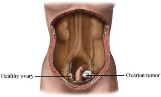 Researchers reported in a study that tiny ovarian tumors exist unobserved in fallopian tubes for an average of four years before they grow large enough to be detected and therefore the diagnosis of those tumors comes too late to save a woman’s life.
The researchers said as the cancer is hard to detect before it has spread, they are finding ways to improve the cancer testing.
Howard Hughes Medical Institute researcher Dr. Patrick Brown of Stanford University in California, who led the study, said in a statement, “Reliable early detection can save more lives than many new anticancer drugs”.
Ovarian cancer kills 140,000 women every year globally and 15,000 in the United States alone. Genetic mutations are known to raise the risk, but most patients do not have a clear genetic risk, and no good screening test exists.
Source: Reuters
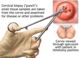
Cone Biopsy – Best Method In Detecting Cervical Disorders!
 Are you suffering with cervical disorder or cervical cancer?
Cone biopsy is the surgery which will help in removing the tumors which are responsible for cervical disorders.
Cervical cancer is the common problem for most of the women. It mainly shows its effect on your reproductive system.
Cone biopsy is an extensive method of cervical biopsy.
In this method, a piece of tissue which is cylindrical in shape is taken for the diagnosis of the cervical cancer. Small tumors in the cervix can be removed with cone biopsy.
Cone biopsy is generally recommended for two main purposes:
- It is used to take the thin or thick sample tissues inside the cervix, which is needed for diagnosis of the cervical cancer.
- It is also used for the treatment of some abnormal tissues or tumors present inside the cervix, which does not need any long term treatment or surgeries to be removed.
Cone biopsy is usually done in either of two given ways:
- LEEP, loop electrosurgical excision procedure is one of the surgical methods of cone biopsy, in which the tissue is removed with the help of an electric wire, heated with an electric current. In this method of surgery, you will be given only with local anesthesia and it is a quick process which can be done at your physician’s office.
- Cone biopsy using laser technology is another method of removing the tumors from the cervix. In this method, CO2 laser is used to remove the abnormal tissues. In this method, you will be given with general anesthesia. Using lasers is always advantageous, as there will be no bleeding process involved when it is done with the lasers.
Important tips to be followed after cone biopsy:
- After the surgery, you need to take rest for one to four hours and also special instructions are given to take care of yourself after the surgery is done.
- You can find some vaginal bleeding for about a week after the surgery, which is quite normal and also some vaginal spotting for up to three weeks.
- Avoid sexual intercourse and use of tampons for four to six weeks.
There are certain complications involved with this cone biopsy. Before going to have this surgery, you need to know about these things which can cause harm to you.
- After surgery, you may need blood transfusion or vaginal packing. This happens very rarely. About only 10% of women or less than that suffer with this problem.
- The cervix can be turned narrow which leads to infertility.
- Due to the lack of ability of the cervix to remain closed during pregnancy, your pregnancy may end up with miscarriages or even premature babies.
Try to discuss with your doctor about these complications before having this surgery in order to avoid the disasters to happen.
Women Who Have Had Breast Cancer Should Avoid Alcohol
 Any woman who has had cancer in one of her breasts is at risk for developing cancer in the second breast.
A new study suggests drinking alcohol may increase this risk of breast cancer by 30 percent.
The longer the women in the study had been users of alcohol, the more likely they were to develop cancer in the second breast.
There are may factors that influence the risk of breast cancer that are beyond women’s control, including gender, age, genetic risk factors, density of breast tissue, onset of menses, late menopause, and family and personal history of breast cancer.
African American women are at greater risk for dying from breast cancer, although more cases of breast cancer occur in white women. Risk factors for breast cancer that women can control are breastfeeding their children, maintaining a healthy weight, exercising regularly, and now, keeping alcohol consumption to a minimum.
The standard screening test for breast cancer is a mammogram. A woman should have her first mammogram procedure at the age of 40 unless there are risk factors that indicate beginning screening earlier. Women with a family history of breast cancer may wish to consider genetic screening as well.
Tuesday, August 11, 2009
HIV, National Women And Girls Awareness Day
 Each year thousands of women worldwide are infected with the HIV virus, this is particularly true amongst black women between the ages 19 to 35, who do not understand the importance of protecting themselves during a sexual relationship with their male partners.
In March, there will be a national day dedicated to HIV and AIDS alertness, organised throughout the U.S. in order to bring more information to all those women who still do not understand how important it is to protect themselves from the virus.
During the National Women and Girls Awareness Day, there will be the possibility to have HIV screening tests and collect as much information on the virus as possible.
It is essential that all women abstain from unsafe sex or use a condom every time they have sex with a partner they do not know well enough.
Unfortunately there are also cases of regular partners who in turn have unsafe sex with other infected people, thus spreading the virus to those who do take precautions; this is why one must be tested regularly.
These tests available to all women will help them find out if they are infected, and if so they may be treated at an early stage and be helped to keep healthy.
It is also important to be aware of your state of health in order not to spread the disease, should you be infected by the virus.
Pregnant women receive the HIV screening test during their first visit at the parental care and during the period of their gestation; this should be so for all women, whether or not they are expecting a baby, they should be offered regular tests as part of a regular health plan.
Monday, July 27, 2009
A to Z List of Cancers
 | |||||||||
A
Acute Lymphoblastic Leukemia, Adult
Acute Lymphoblastic Leukemia, Childhood
Acute Myeloid Leukemia, Adult
Acute Myeloid Leukemia, Childhood
Adrenocortical Carcinoma
Adrenocortical Carcinoma, Childhood
AIDS-Related Cancers
AIDS-Related Lymphoma
Anal Cancer
Appendix Cancer
Astrocytoma, Childhood Cerebellar
Astrocytoma, Childhood Cerebral
Atypical Teratoid/Rhabdoid Tumor, Childhood, Central Nervous System
B
Basal Cell Carcinoma, see Skin Cancer (Nonmelanoma)Bile Duct Cancer, Extrahepatic
Bladder Cancer
Bladder Cancer, Childhood
Bone Cancer, Osteosarcoma and Malignant Fibrous Histiocytoma
Brain Stem Glioma, Childhood
Brain Tumor, Adult
Brain Tumor, Brain Stem Glioma, Childhood
Brain Tumor, Central Nervous System Atypical Teratoid/Rhabdoid Tumor, Childhood
Brain Tumor, Central Nervous System Embryonal Tumors, Childhood
(See What Are Childhood Central Nervous System Embryonal Tumors?)
Brain Tumor, Cerebellar Astrocytoma, Childhood
Brain Tumor, Cerebral Astrocytoma/Malignant Glioma, Childhood
Brain Tumor, Craniopharyngioma, Childhood
Brain Tumor, Ependymoblastoma, Childhood
(See What Are Childhood Central Nervous System Embryonal Tumors?)
Brain Tumor, Ependymoma, Childhood
Brain Tumor, Medulloblastoma, Childhood
Brain Tumor, Medulloepithelioma, Childhood
(See What Are Childhood Central Nervous System Embryonal Tumors?)
Brain Tumor, Pineal Parenchymal Tumors of Intermediate Differentiation, Childhood
(See What Are Childhood Central Nervous System Embryonal Tumors?)
Brain Tumor, Supratentorial Primitive Neuroectodermal Tumors and Pineoblastoma, Childhood
Brain Tumor, Visual Pathway and Hypothalamic Glioma, Childhood
Brain and Spinal Cord Tumors, Childhood (Other)
Breast Cancer
Breast Cancer and Pregnancy
Breast Cancer, Childhood
Breast Cancer, Male
Bronchial Tumors, Childhood
Burkitt Lymphoma
C
Carcinoid Tumor, Childhood
Carcinoid Tumor,Gastrointestinal
Carcinoma of Unknown Primary
Central Nervous System Atypical Teratoid/Rhabdoid Tumor, Childhood
Central Nervous System Embryonal Tumors, Childhood
(See What Are Childhood Central Nervous System Embryonal Tumors?)
Central Nervous System Lymphoma, Primary
Cerebellar Astrocytoma, Childhood
Cerebral Astrocytoma/Malignant Glioma, Childhood
Cervical Cancer
Cervical Cancer, Childhood
Childhood Cancers
Chordoma, Childhood
Chronic Lymphocytic Leukemia
Chronic Myelogenous Leukemia
Chronic Myeloproliferative Disorders
Colon Cancer
Colorectal Cancer, Childhood
Craniopharyngioma, Childhood
Cutaneous T-Cell Lymphoma, see Mycosis Fungoides and Sézary Syndrome
D
[No Entries]
E
Embryonal Tumors, Central Nervous System, Childhood
(See What Are Childhood Central Nervous System Embryonal Tumors?)
Endometrial Cancer
Ependymoblastoma, Childhood
(See What Are Childhood Central Nervous System Embryonal Tumors?)
Ependymoma, Childhood
Esophageal Cancer
Esophageal Cancer, Childhood
Ewing Family of Tumors
Extracranial Germ Cell Tumor, Childhood
Extragonadal Germ Cell Tumor
Extrahepatic Bile Duct Cancer
Eye Cancer, Intraocular Melanoma
Eye Cancer, Retinoblastoma
F
[No Entries]
G
Gallbladder Cancer
Gastric (Stomach) Cancer
Gastric (Stomach) Cancer, Childhood
Gastrointestinal Carcinoid Tumor
Gastrointestinal Stromal Tumor (GIST)
Gastrointestinal Stromal Cell Tumor, Childhood
Germ Cell Tumor, Extracranial, Childhood
Germ Cell Tumor, Extragonadal
Germ Cell Tumor, Ovarian
Gestational Trophoblastic Tumor
Glioma, Adult
Glioma, Childhood Brain Stem
Glioma, Childhood Cerebral Astrocytoma
Glioma, Childhood Visual Pathway and Hypothalamic
H
Hairy Cell Leukemia
Head and Neck Cancer
Hepatocellular (Liver) Cancer, Adult (Primary)
Hepatocellular (Liver) Cancer, Childhood (Primary)
Histiocytosis, Langerhans Cell
Hodgkin Lymphoma, Adult
Hodgkin Lymphoma, Childhood
Hypopharyngeal Cancer
Hypothalamic and Visual Pathway Glioma, Childhood
I
Intraocular Melanoma
Islet Cell Tumors (Endocrine Pancreas)
J
[No Entries]
K
Kaposi Sarcoma
Kidney (Renal Cell) Cancer
Kidney Cancer, Childhood
L
Langerhans Cell Histiocytosis
Laryngeal Cancer
Laryngeal Cancer, Childhood
Leukemia, Acute Lymphoblastic, Adult
Leukemia, Acute Lymphoblastic, Childhood
Leukemia, Acute Myeloid, Adult
Leukemia, Acute Myeloid, Childhood
Leukemia, Chronic Lymphocytic
Leukemia, Chronic Myelogenous
Leukemia, Hairy Cell
Lip and Oral Cavity Cancer
Liver Cancer, Adult (Primary)
Liver Cancer, Childhood (Primary)
Lung Cancer, Non-Small Cell
Lung Cancer, Small Cell
Lymphoma, AIDS-Related
Lymphoma, Burkitt
Lymphoma, Cutaneous T-Cell, see Mycosis Fungoides and Sézary Syndrome
Lymphoma, Hodgkin, Adult
Lymphoma, Hodgkin, Childhood
Lymphoma, Non-Hodgkin, Adult
Lymphoma, Non-Hodgkin, Childhood
Lymphoma, Primary Central Nervous System
M
Macroglobulinemia, Waldenström
Malignant Fibrous Histiocytoma of Bone and Osteosarcoma
Medulloblastoma, Childhood
Medulloepithelioma, Childhood
(See What Are Childhood Central Nervous System Embryonal Tumors?)
Melanoma
Melanoma, Intraocular (Eye)
Merkel Cell Carcinoma
Mesothelioma, Adult Malignant
Mesothelioma, Childhood
Metastatic Squamous Neck Cancer with Occult Primary
Mouth Cancer
Multiple Endocrine Neoplasia Syndrome, Childhood
Multiple Myeloma/Plasma Cell Neoplasm
Mycosis Fungoides
Myelodysplastic Syndromes
Myelodysplastic/Myeloproliferative Diseases
Myelogenous Leukemia, Chronic
Myeloid Leukemia, Adult Acute
Myeloid Leukemia, Childhood Acute
Myeloma, Multiple
Myeloproliferative Disorders, Chronic
N
Nasal Cavity and Paranasal Sinus Cancer
Nasopharyngeal Cancer
Nasopharyngeal Cancer, Childhood
Neuroblastoma
Non-Hodgkin Lymphoma, Adult
Non-Hodgkin Lymphoma, Childhood
Non-Small Cell Lung Cancer
O
Oral Cancer, Childhood
Oral Cavity Cancer, Lip and
Oropharyngeal Cancer
Osteosarcoma and Malignant Fibrous Histiocytoma of Bone
Ovarian Cancer, Childhood
Ovarian Epithelial Cancer
Ovarian Germ Cell Tumor
Ovarian Low Malignant Potential Tumor
P
Pancreatic Cancer
Pancreatic Cancer, Childhood
Pancreatic Cancer, Islet Cell Tumors
Papillomatosis, Childhood
Paranasal Sinus and Nasal Cavity Cancer
Parathyroid Cancer
Penile Cancer
Pharyngeal Cancer
Pheochromocytoma
Pineal Parenchymal Tumors of Intermediate Differentiation, Childhood
(See What Are Childhood Central Nervous System Embryonal Tumors?)
Pineoblastoma and Supratentorial Primitive Neuroectodermal Tumors, Childhood
Pituitary Tumor
Plasma Cell Neoplasm/Multiple Myeloma
Pleuropulmonary Blastoma
Pregnancy and Breast Cancer
Primary Central Nervous System Lymphoma
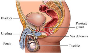
Prostate Cancer
Q
[No Entries]
R
Rectal Cancer
Renal Cell (Kidney) Cancer
Renal Cell (Kidney) Cancer, Childhood
Renal Pelvis and Ureter, Transitional Cell Cancer
Respiratory Tract Carcinoma Involving the NUT Gene on Chromosome 15
Retinoblastoma
Rhabdomyosarcoma, Childhood
S
Salivary Gland Cancer
Salivary Gland Cancer, Childhood
Sarcoma, Ewing Family of Tumors
Sarcoma, Kaposi
Sarcoma, Soft Tissue, Adult
Sarcoma, Soft Tissue, Childhood
Sarcoma, Uterine
Sézary Syndrome
Skin Cancer (Nonmelanoma)
Skin Cancer, Childhood
Skin Cancer (Melanoma)
Skin Carcinoma, Merkel Cell
Small Cell Lung Cancer
Small Intestine Cancer
Soft Tissue Sarcoma, Adult
Soft Tissue Sarcoma, Childhood
Squamous Cell Carcinoma, see Skin Cancer (Nonmelanoma)
Squamous Neck Cancer with Occult Primary, Metastatic
Stomach (Gastric) Cancer
Stomach (Gastric) Cancer, Childhood
Supratentorial Primitive Neuroectodermal Tumors, Childhood
T
T-Cell Lymphoma, Cutaneous, see Mycosis Fungoides and Sézary Syndrome
Testicular Cancer
Throat Cancer
Thymoma and Thymic Carcinoma
Thymoma and Thymic Carcinoma, Childhood
Thyroid Cancer
Thyroid Cancer, Childhood
Transitional Cell Cancer of the Renal Pelvis and Ureter
Trophoblastic Tumor, Gestational
U
Unknown Primary Site, Carcinoma of, Adult
Unknown Primary Site, Cancer of, Childhood
Unusual Cancers of Childhood
Ureter and Renal Pelvis, Transitional Cell Cancer
Urethral Cancer
Uterine Cancer, Endometrial
Uterine Sarcoma
V
Vaginal Cancer
Vaginal Cancer, Childhood
Visual Pathway and Hypothalamic Glioma, Childhood
Vulvar Cancer
W
Waldenström Macroglobulinemia
Wilms Tumor
Women's Cancers
X
[No Entries]
Y
[No Entries]
Z
[No Entries]
(source: http://www.nci.nih.go)
Subscribe to:
Comments (Atom)

